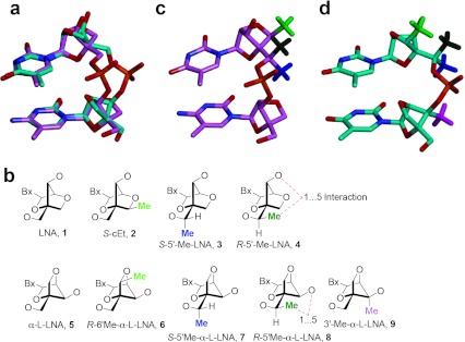
Figure 6.
Structural models of LNA and α-L-LNA dinucleotide units showing relative orientations of 3′-Me, 5′-Me, and 6′-Me groups. (a) Overlay of common structural units such as nucleobases, C1′ carbon and O4′ oxygen atoms of LNA (pink carbons) and α-L-LNA (teal carbons) dimers obtained from the NMR structures of modified DNA/RNA duplexes (refs. 32 and 15). Complementary RNA strand not shown for clarity. (b) Structures of the monomeric units showing relative orientations of various methyl groups. (c) Relative orientations of S-6′-Me (light green), R-5′-Me (olive green), and S-5′-Me (dark blue) groups in a LNA dinucleotide unit. (d) Relative orientations of R-6′-Me (light green), R-5′-Me (olive green), S-5′-Me (dark blue), and 3′-Me (pink) groups in an α-L-LNA dinucleotide unit. LNA, locked nucleic acid.

