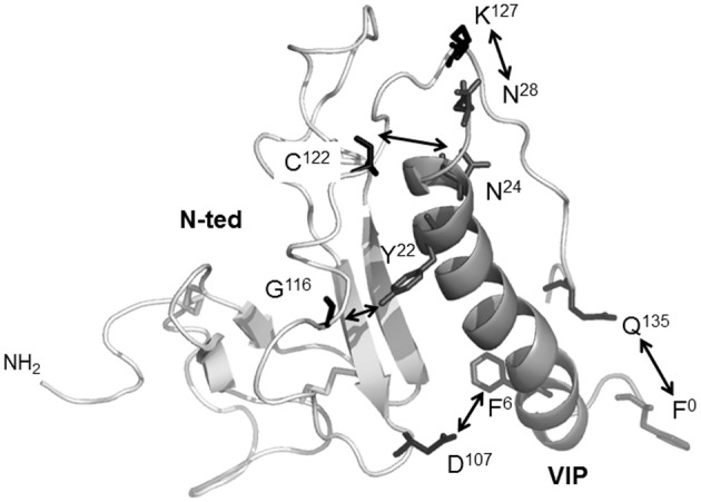
Figure 3.

The 3D-structural model of VPAC1 receptor N-ted and docking of VIP. Ribbon representation of the VPAC1 N-ted: light gray ribbon, main chain; white ribbon, VIP. Docking calculations showed that Q135, D107, G116, C122, and K127 residues (middle gray sticks) present in the N-ted were in contact (white arrows) with the side chains of F0, F6, Y22, N24, and N28 (black sticks) of VIP residues, respectively. Figure was obtained by using PyMOL software (http://www.pymol.org).
