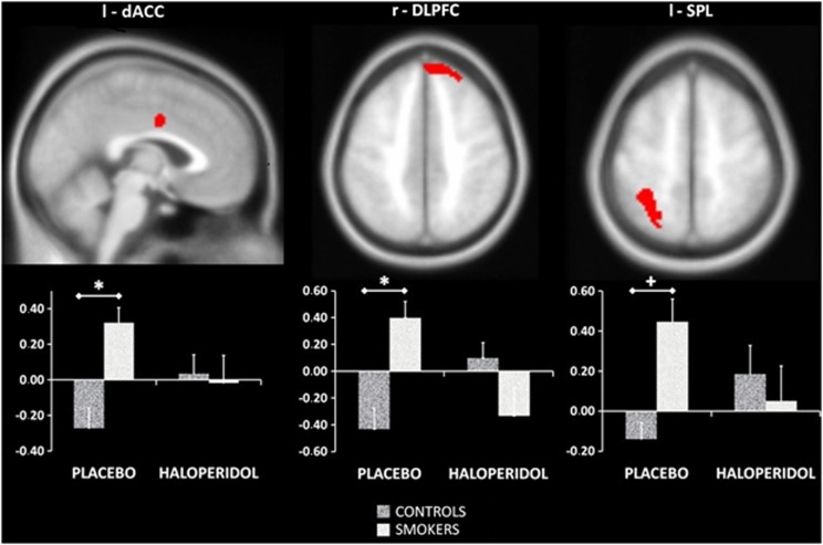
Figure 3.
Group differences for brain activation associated with attentional bias. *p<0.05 FWE small volume corrected both with and without masking for the group × medication interaction, +p<0.05 FWE small volume corrected only without masking for the group × medication interaction. The values on the Y axis represent contrast values for LCSP minus LCNP, consequently positive values on the Y axis indicate more brain activation for line counting when smoking-related pictures are presented on the background relative to when neutral pictures are presented on the background. FWE, family wise error; LCNP, line-counting neutral picture; LCSP, line-counting smoke picture; l, left; r, right; dACC, dorsal anterior cingulate gyrus; DLPFC, dorsolateral prefrontal cortex; SPL, superior parietal lobe.

