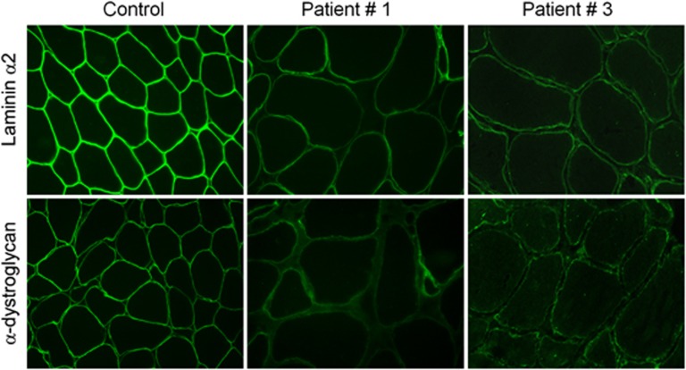
Figure 1.
Reduced α-DG glycosylation in POMT1-mutated patients. α-DG immunostaining using an antibody directed against a glycosylated epitope shows a normal labeling at the periphery of each fiber in the control's muscle, in comparison with patients 1 and 3 where the majority of myofibers show a faint immunoreaction and variability of the intensity of the labeling. Laminin α2 immunostaining using an antibody directed against the 80-kDa carboxyl-terminus shows a subtle reduction of the labeling in the patients.

