
Figure 5.
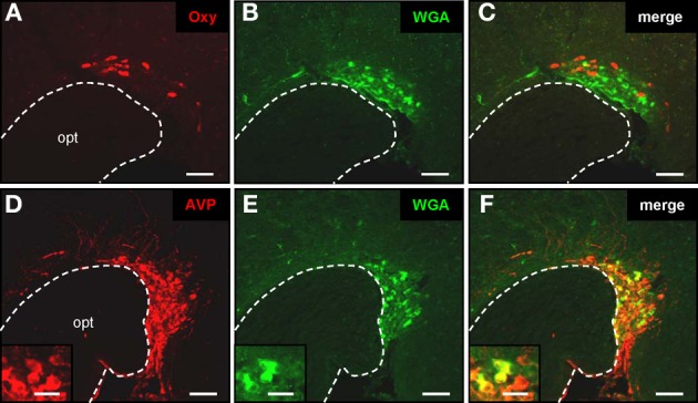
Transsynaptic labeling of vasopressinergic neurons in the supraoptic nucleus. (A–C) Cross section through the supraoptic nucleus. By means of a specific anti-oxytocin antibody, multiple oxytonergic neurons can be visualized near the optic tract (opt) (A; red). On the same section WGA-immunoreactive cells can be observed (B; green). Merging the two pictures (C) reveals that oxytonergic neurons are not labeled by the transsynaptic marker. Scale bars: 50 μm. (D–F) Tissue section through the supraoptic nucleus. After immunohistochemical staining with an anti-neurophysin-vasopressin antibody, neurons are visible in close vicinity to the optic tract (opt) (A; red). On the same section WGA-immunoreactive cells are located (B; green). Merge of pictures (A) and (B) reveals that vasopressinergic cells are labeled by the transsynaptic marker (C; yellow). Scale bars: 50 μm. Higher magnifications are shown on the insets. Scale bars: 20 μm. All pictures represent wide field images.