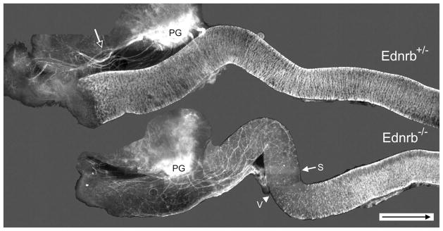
Figure 1.
Distribution of YFP expression in freshly isolated E16.5 Ednrb+/− and Ednrb−/− hindgut. In the Ednrb+/− colon the majority of nerve fibers (open arrow) radiate from the pelvic ganglia (PG) and travel outside the colon until reaching the anal region; the entire colon is occupied by ganglia shown as white lines. In comparison, most fibers enter the Ednrb−/− distal colon and project in oral and aboral directions. Arrow indicates the most oral position of sacral nerve fibers; arrowhead indicates the most aboral position of the vagal crest wavefront. The figure is a photomontage. Arrow inside scale bar indicates oral direction. Scale bar = 500 μm.

