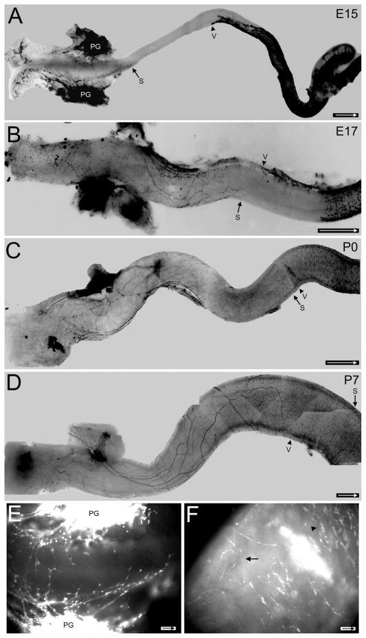
Figure 2.
Development of sacral fibers in freshly isolated Ednrb−/− colons. In A–D arrow indicates the most oral position of the sacral wavefront, arrowhead shows the most aboral position of the vagal wavefront. YFP signals are dark in A–D and light in E,F. A: At E15 processes from the pelvic ganglion project in oral and aboral directions. The vagal wavefront has moved into the midcolon and will not advance much further. B: At E17 nerve fibers have advanced orally and form a network of large and small processes. The vagal wavefront is found at the midcolon with the mesenteric strand extending aborally and from it crest cells will move laterally across the width of the colon. C: At birth (P0) the sacral nerve fibers have advanced to the site of the vagal wavefront. Note that the vagal wavefront contains many cells. D: At P7 large sacral fibers are apparent and some extend into the region occupied by the vagal crest-derived cells while smaller fibers extend extensive processes of varying sizes. The sacral fibers and vagal crest have also begun to intermingle at the midcolon. E: An enlargement of the terminal region of (A). Nerve fibers arise from pelvic ganglia (PG). Note the enlarged swellings are sacral-derived neural crest cells along the nerve fibers. F: An enlargement of the midcolon where the sacral fibers (arrow) and vagal crest cell wavefront (arrowhead) meet in the P0 colon in (C). A–D are photomontages. Arrow inside scale bar indicates oral direction. Scale bars = 500 μm in A; 250 μm in B; 1 mm in C,D; 50 μm in E; 100 μm in F.

