Figure 3.
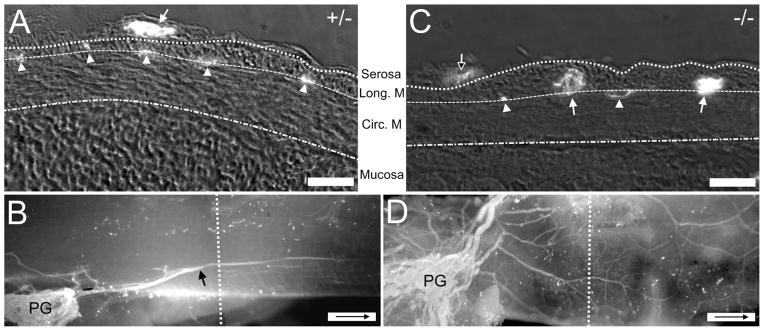
Comparison of the location of nerve fibers with respect to the wall of the colon in Ednrb+/− and Ednrb−/− preparations. A: Cross-section of P9 Ednrb+/− colon. Fluorescence indicates the presence of neurofilament-IR (bright spots, NF). Arrow indicates large sacral fascicle superficial to longitudinal muscle. Arrowhead shows fibers within the muscle layers indicating the location of the myenteric plexus. B: A montage showing a whole-mount view of the colon sectioned in (A). An extrinsic sacral fiber (arrow) arises from the pelvic ganglia (PG). The dotted line indicates the approximate area where the cross-section was cut. C: Cross-section of P9 Ednrb−/− colon. Large NF+ sacral fiber bundles (arrows) and smaller fiber bundles (arrowheads) are located between the muscle layers except for one small fascicle (open arrow) on the surface of the longitudinal muscle. D: A montage showing a whole-mount view of the colon sectioned in (C). Multiple fiber bundles are present although ganglia are not. The dotted line shows the position where section (C) was cut. Arrow inside scale bar indicates oral direction. Scale bars = 30 μm in A,C; 500 μm in B,D.

