Figure 4.

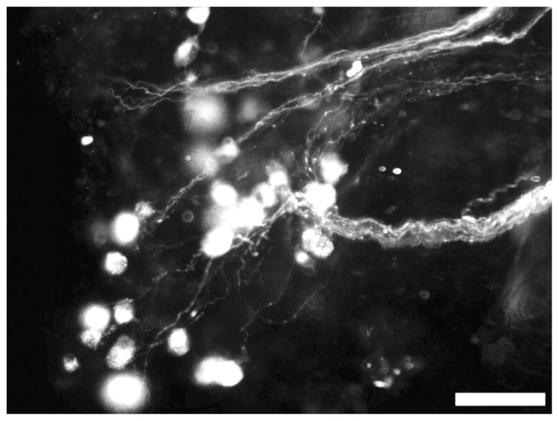
Cells in the pelvic ganglion from a P29 Ednrb−/− mouse are fluorescent after application of DiI crystals to nerve fibers in the mid-colon. Scale bar = 50 μm.

Cells in the pelvic ganglion from a P29 Ednrb−/− mouse are fluorescent after application of DiI crystals to nerve fibers in the mid-colon. Scale bar = 50 μm.