Figure 6.
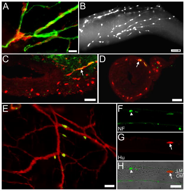
Cells on nerve fibers in Ednrb−/− preparations. A: Surface view. E14.5 YFP+ SNCCs (red) appear along βIII-tubulin-immunoreactive nerve fibers (green) within the distal colon. B: Surface view. E15.5 YFP+ cells are found along nerve fibers in the distal colon heading in an oral direction (arrow inside scale bar indicates oral direction). C: Section through another E15.5 distal colon just oral to the pelvic ganglion. A βlll-tubulin-immunoreactive fiber (green) and YFP+ cells (red) together enter the wall of the colon where many YFP+ cells are already present. D: Section through the E15.5 distal colon in a position more oral than (C). A YFP+ cell (red) and βlll-tubulin-immunoreactive fibers (green) are colocalized (arrow); numerous YFP+ cells are found between the muscle layers. E: Whole mount of P7 distal colon. Hu+ (green) cells are found on YFP+ (red) nerve fibers near the pelvic ganglia. F–H: Cross-section of P31 distal colon. (F) Both a faintly stained YFP+ cell and neurofilament-immunoreactive nerve fibers (green, arrowhead) are located between the muscle layers while nerve fibers are also found at the edge of the muscle-submucosa interface. The submucosa and the mucosa were removed. (G) An Hu+ (red) neuron is found between the muscle layers usually occupied by the myenteric ganglia. (H) Overlay of images with brightfield image and the circular (CM) and longitudinal muscles (LM) labeled. Scale bars = 50 μm in A,C–H; 100 μm in B. A magenta-green copy of this figure is available as Supporting Figure 3.

