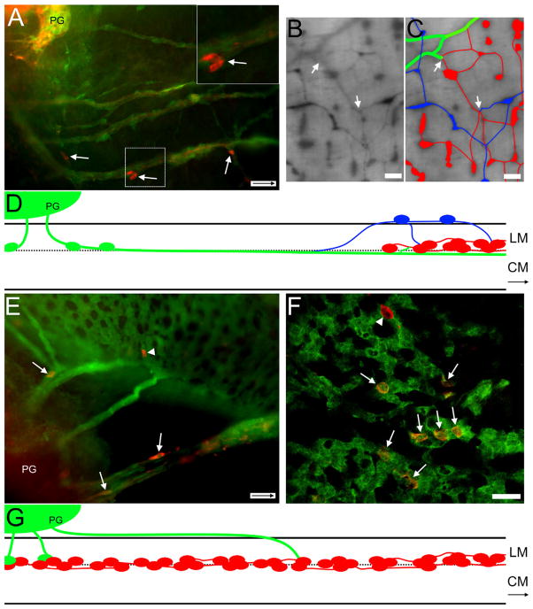
Figure 7.
A: E16.5 Ednrb−/−. TH+ cells (arrows) and fibers accompany sacral fibers projecting orally from the PG into the colon. Note the absence of ganglia. Inset, upper right: Two TH+ cells within box are shown at higher magnification. B: P27 Ednrb−/−. Changes in position of extrinsic fibers when encountering intrinsic ganglia. Neural crest-derived cells are labeled with β-galactosidase. Cells and fibers on the serosal surface are sharp and dark while cells and fibers within the muscle layers are lighter and blurry. C: Overlay of fibers shown in (B). At the site where the sacral fibers extend into the region of vagal ganglia in the midcolon, large sacral fibers (green) are located between the muscle layers, the same level as vagal ganglia (red). There are also ganglia in the serosal region (blue), above the vagal ganglia, whose origin could be either cells that traveled from sacral axons and/or from the vagally derived intrinsic neurons in the hypoganglionic region. These different elements appear to intersect (arrows). D: Diagram summarizing the findings for Ednrb−/− aganglionic colon. Sacral extrinsic fibers and cells (green) projecting from the pelvic ganglia (PG) intersect with the vagal myenteric plexus (red) in the mid-colon. A population of cells and smaller fascicles (blue) appear at the vagal wavefront. They exit the myenteric locale and reside in the serosal region superficial to the longitudinal muscle (LM) where they appear to have connections with vagal ganglia. The larger sacral fascicles continue orally between the muscle layers where they appear to intermingle with the vagal ganglia. CM: circular muscle. E: Ednrb+/−. YFP+ sacral fibers (green) extending from the pelvic ganglia (PG) into the distal colon contain TH+ cells (red, arrows). A single TH+ cell (arrowhead) is incorporated into ganglia formed by VNCCs. F: E16.5 Ednrb+/− colon. A confocal image showing TH+ (red, arrows) YFP+ SNCCs among the YFP+ VNCCs (green) forming nascent ganglia. Note a single TH+ cell that is not YFP+ (arrowhead). G: Diagram summarizing the findings for Ednrb+/− ganglionated colon. Fibers leaving the pelvic ganglion travel the longest distance superficial to the longitudinal muscle before penetrating the muscle and terminating between the muscle layers. SNCC (green) cells move along extrinsic fibers to reach VNCC myenteric ganglia (red) where they become incorporated into the ganglia. Arrows inside scale bar indicate oral direction. Scale bars = 50 μm in A,E; 250 μm in B,C; 25 μm in F; D,G not to scale. A magenta-green copy of this figure is available as Supporting Figure 4.

