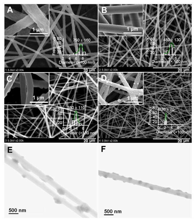
Figure 3.
SEM images of PAN nanofibers prepared using different processes: (A) surface morphologies and diameter distribution of nanofibers F1 from single fluid electrospinning; (B) surface morphologies and diameter distribution of nanofibers F2 from a modified coaxial process with only DMAc as sheath fluid; (C and D) surface morphologies and diameter distribution of nanofibers F3 and F4 from a modified coaxial process with AgNO3 solution as sheath fluid, respectively. The scale bars in the insets of (C and D) represent 500 nm. (E and F) are TEM images of nanofibers F3 and F4, respectively.
Abbreviations: AgNO3, silver nitrate; DMAc, N,N-dimethylacetamide; PAN, polyacrylonitrile; SEM, scanning electron microscopy; TEM, transmission electron microscopy.

