Figure 5.

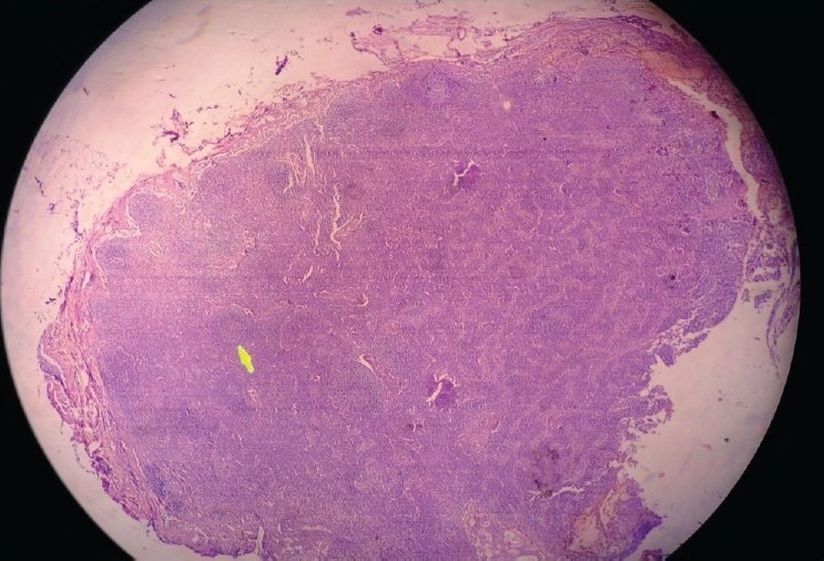
Low magnification microscopy of an axillary lymph node showing effacement of lymph node architecture with a preserved lymphoid follicle (arrow) (H and E, 10×)

Low magnification microscopy of an axillary lymph node showing effacement of lymph node architecture with a preserved lymphoid follicle (arrow) (H and E, 10×)