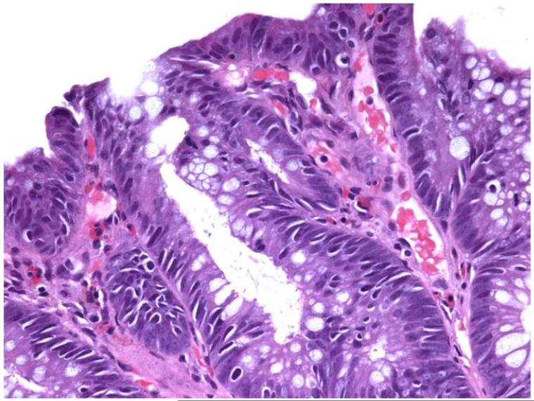
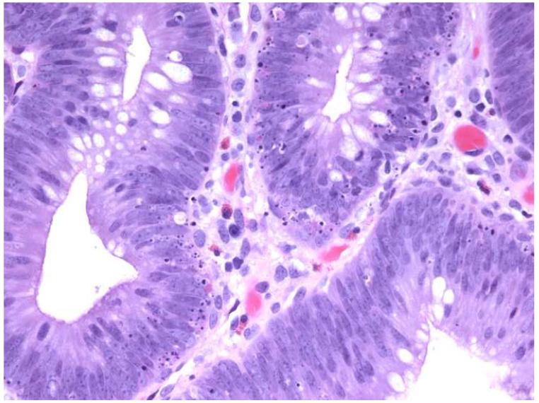
Figure 1.
Photomicrographs of hematoxylin and eosin-stained tissue sections from an adenoma in an HNPCC patient (a) showing high numbers of AILs (>10/HPF) and low numbers of intra-adenomatous apoptoses (0-4/HPF) and a control adenoma (b) with low numbers of AILs (0-4/HPF) and high numbers of intra-adenomatous apoptoses (>10/HPF) (original magnification: 400x).


