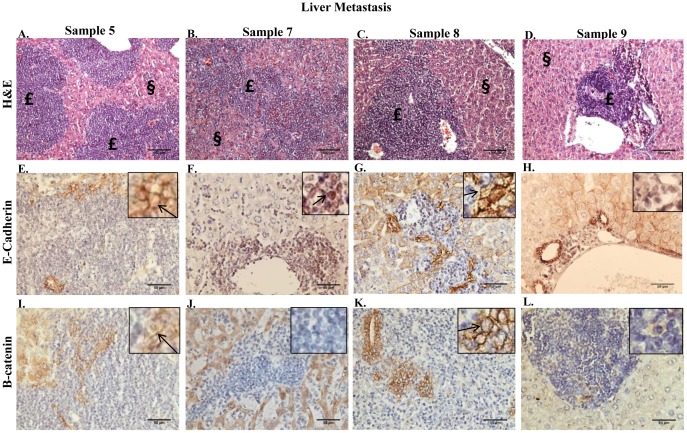
Figure 4. Metastatic lesions in the livers of mice bearing mammary fat pad tumors that were derived from transplantation of bone marrow aspirate containing metastatic tumor cells.
A–D. H+E staining performed on 5 µm paraffin-embedded sections of liver from sample 5–7, and 9 BM illustrates metastatic lesions. Lesion indicated by £; normal tissue indicated by §. 100× magnification. E–H. IHC performed on 5 µm paraffin-embedded sections of liver from sample 5–7, and 9 BM using a monoclonal antibody to E-cadherin. I–L. IHC performed on 5 µm paraffin-embedded sections of liver from sample 5–7, and 9 BM using a polyclonal antibody to β-catenin.

