
Figure 1.
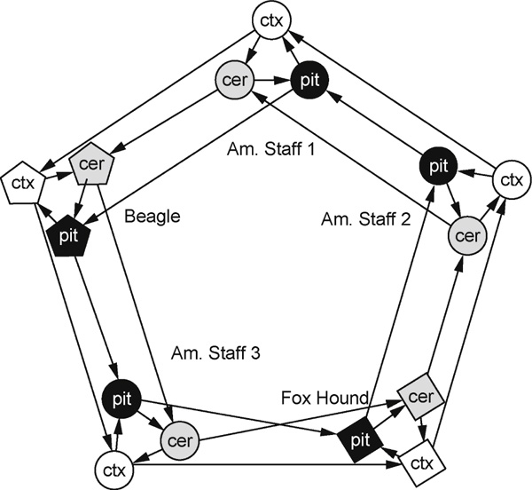
Experimental design, consisting of five loops contrasting each of the three brain tissues [cortex (ctx), cerebellum (cer) and pituitary (pit)] from a single dog, nested within three loops contrasting the same tissue across each of the five dogs. Arrowheads point to the Cy5 sample on each array, and arrow bases lead from the Cy3 sample. Each tissue in each dog is represented by four hybridisations with a balance of dye flips.