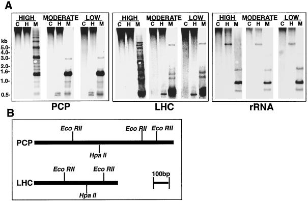
Figure 3.
A. carterae PCP and LHC loci are normally hypermethylated at CpG motifs and their methylation status is modified by light conditions. A, Southern analysis of PCP, LHC, and rDNA loci in cells grown under different light conditions using the restriction enzymes HpaII (H) and MspI (M); C, controls. The sizes indicated to the left of the figure correspond to positions of DNA size standards. Note that the apparently repeating pattern seen for the MspI digest of high-light-grown cells (A, left panel) is consistent with the hypothesis that several of the PCP loci are tandemly organized (Sharples et al., 1996). B, Restriction maps of single A. carterae PCP (based on figure 6A of Sharples et al., 1996; bp 35–1110)- and LHC (based on figure 1B of Hiller et al., 1995, bp 10–600)-coding sequences that were used as probes.

