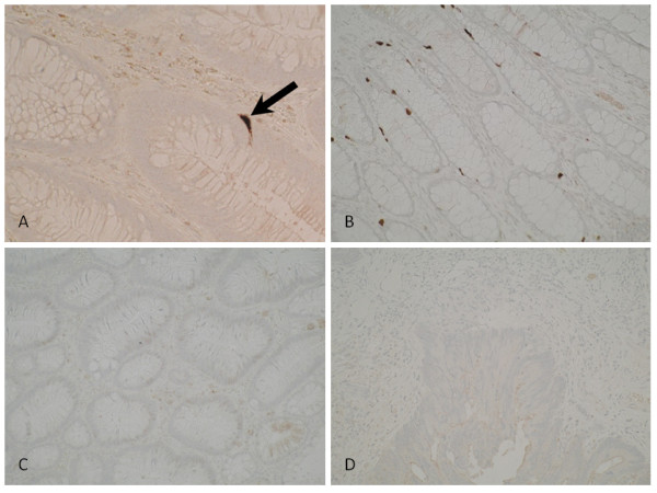
Figure 1.

Photomicrographs of colonic tissue immunohistochemically stained for PSCA. Intense staining can be seen in a crypt neuroendocrine cell (1A, arrow). Colonocytes show little or no staining across all stages of the adenoma-carcinoma sequence. No changes in intensity or topographic distribution of PSCA expression were observed between normal mucosa (1B), adenomatous tissue displaying low grade epithelial dysplasia (1C), and invasive carcinoma (1D).
