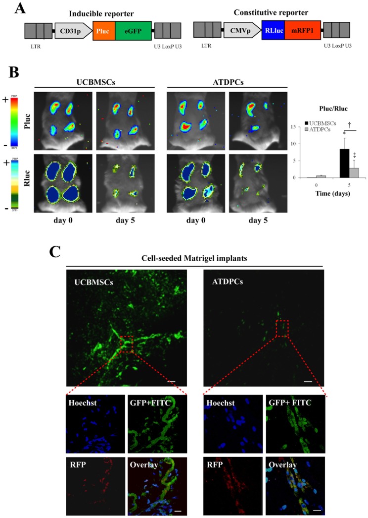
Figure 4. Participation of UCBMSCs in functional microvascular structures in vivo.
A) Schematic representations of the inducible (CD31p-Pluc-eGFP) and constitutive (CMVp-Rluc-RFP) lentiviral vectors, in which each luciferase and fluorescent signal generated following promoter activation appears remarked with the corresponding colour. B) Representative registrations of Pluc and Rluc activities from dual labelled UCBMSCs and ATDPCs co-implanted with Matrigel superimposed on black white dorsal images of the recipient animal. Colour bars illustrating relative light intensities from Pluc and Rluc; low: blue and black; high: red and blue, respectively. Histogram represents over time quantification of implanted cell differentiation degree measured as the ratio between Pluc and Rluc signals. The region-of-interest (ROI) for the quantification of photon emission included only the individual injection sites. *P<0.001, † P = 0.032 and ‡ P = 0.047. C) Representative green-fluorescence images showing formation of microcirculatory vessels within UCBMSC- and ATDPC-seeded Matrigel implants removed from animals in which circulatory system was previously filled with FITC-dextran. Bars = 100 µm. Additional merged images show a functionally connected cell network better organized within Matrigel implants seeded with fluorescent labelled UCBMSCs than in those seeded with ATDPCs. ATDPCs appear more disperse and not well-organized. Bars = 20 µm.

