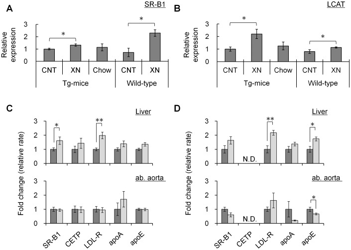
Figure 4. Expression analyses in mice liver and ab. aorta.
(A) SR-B1 and (B) LCAT protein expression in liver. Data was standardized for β-actin expression. (N = 15; CETP-Tg mice control, N = 18; CETP-Tg mice xanthohumol, N = 11; CETP-Tg mice Chow, N = 6; wild-type mice control, N = 8; wild-type mice xanthohumol) (C, D) Transcript analyses of liver (upper) and ab. aorta (lower) in CETP-Tg mice (C) and wild-type mice (D). (N = 12; CETP-Tg mice control, N = 13; CETP-Tg mice xanthohumol, N = 9 to 10; CETP-Tg mice Chow, N = 5; wild-type mice control, N = 7 to 8; wild-type mice xanthohumol) All data were standardized for GAPDH expression. Expression levels of control group (without xanthohumol) were set at 1.0. Means±SEM. *P<0.05, **P<0.01.

