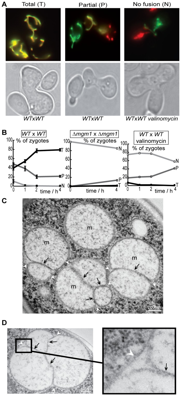
Figure 1. Mitochondrial fusion is inhibited upon dissipation of the mitochondrial membrane potential.

ΔΨm. Wild-type (WT) or Δmgm1 cells expressing red or green fluorescent proteins targeted to the matrix (mtGFP, mtRFP) were conjugated and incubated for 4 h under control conditions or in the presence of valinomycin. A: Fluorescence and phase-contrast microscopy depicts yeast zygotes with total fusion (T: all mitochondria are doubly labeled), partial fusion (P: doubly and simply labeled mitochondria coexist) or no fusion (N: all mitochondria are simply labeled). B: The percentage of zygotes with total (T), partial (P) or no fusion (N) as a function of time. Fusion is inhibited in the absence of Mgm1 or in the presence of valinomycin. C, D: Electron microscopy of valinomycin-treated cells reveals mitochondria with fused outer membranes (white arrowheads) and elongated, aligned inner membranes (black arrows: septae).
