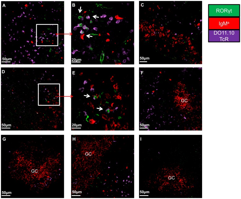
Figure 8. Th17 cells interact with cognate B cells.
At days 3, 7 and 10 post immunisation dLNs were snapped frozen and were analysed for the presence of transgenic Th17 cells by the co-expression of the clonotypic DO11.10 TcR (PURPLE) and RORγt (GREEN). In addition transgenic B cells were detected by staining against IgMa (RED). Representative pictures of dLN sections of: A and B) Th17 recipients 3 days post immunisation C) Th1 recipients 3 days post immunisation D and E) Th17 recipients 7 days post immunisation F) GC-like structures in Th17 recipient 7 days post immunisation G) GC-like structures in Th1 recipient 7 days post immunisation H) GC-like structures in Th17 recipient 10 days post immunisation I) GC-like structure in Th1 recipient 10 days post immunisation. Microscope: Carl Zeiss LSM510 META Confocal, Objectives: B) Zeiss planapochromat 40×/1 NA water objective; A, C–I) Zeiss PH 25×/0.85 NA water objective. Images were acquired using Zeiss LSM510 operating software and off-line image analysis (contrast enhancement and noise removal) were performed using Volocity® software. At least 3 random sections were imaged per mice and at least 4 pictures were acquired per section. Similar results were obtained in one additional experiment.

