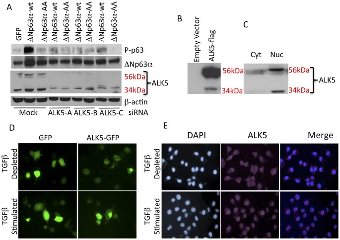
Figure 4. Nuclear accumulation of the intracellular kinase domain of ALK5 (ALK5IKD) in response to TGFβ.
A. Three different siRNAs targeted against ALK5 were transfected into IMEC cells, 48 hrs later cells were transfected with GFP, ΔNp63α-WT and ΔNp63α-AA expression vectors, whole cell lysates were harvested after 24 hrs and analyzed for phospho-p63, total p63, TGFβR1 and β-actin. B. H1299 cells were transfected with an ALK5-WT-Flag expression vector, whole cell lysates were harvested and analyzed for full length and cleaved fragments with anti-Flag antibody. C. Analysis of ALK5 distribution in IMECs indicates that the 34 kDa C-terminal ALK5 fragment is present only in the nucleus. D. H1299 cells were transfected with ALK5-GFP expression vector, 6 hrs after transfection cells were treated with vehicle control or TGFβ1 for 8 hrs and imaged for subcellular distribution of GFP. E. H1299 cells were serum starved for 12 hr and then induced with vehicle control or TGFβ1 for 1hour. Cells were stained with anti- TGFβR1 (V-22) antibody. Depletion of TGFβR1in H1299cells by siRNA is shown as a control for the specificity of the antibody.

