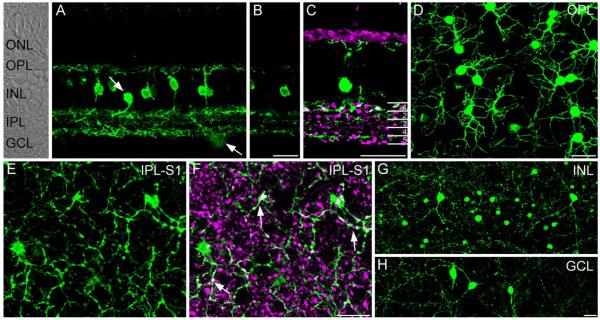
Figure 1.
Localization of SCGN in the peripheral macaque retina. A: Confocal projection (7 ×0.8 μm) of a vertical section of macaque retina stained for SCGN. Labeling is present in the soma, dendrites, and axons of a population of bipolar cells and in occasional putative amacrine cells in the inner nuclear layer and ganglion cell layer (arrows). Retinal layers are indicated on the transmitted light image to the left side of panel A. B: Single confocal plane of the same area shown in A. Note that SCGN-positive processes are concentrated in three major bands at the top, middle, and bottom of the IPL. C: Confocal projection (4 ×0.9 μm) of vertical section stained with vGluT1 and SCGN shows that some SCGN-positive processes in S1 were also immunoreactive for vGluT1, while processes outside of S1 lacked vGluT1 immunoreactivity. Numbers on the right-hand side of panel delineate the five IPL strata. D: Confocal projection of wholemount retina (5 × 0.4 μm) stained for SCGN and focused at the level of the OPL. Strong SCGN immunoreactivity is present in diffuse bipolar cell dendrites. E: Confocal projection (3 ×0.4 μm) of wholemount retina stained for SCGN and focused at the level of S1 of the IPL. F: Same region as in E showing staining for SCGN and vGluT1. SCGN-positive bipolar cell terminals show vGluT1 immunoreactivity (white, arrows). These terminals are fine, with intermittent swellings arranged like “pearls on a string.” Other vGluT1-positive bipolar cell terminals are more bulbous in appearance. Note that many SCGN-stained processes in this focal plane are not vGluT1-positive, and are likely of amacrine cell origin. G,H: Confocal projections (5 ×0.9 μm), showing wide-field amacrine cells with somata located in the inner nuclear layer (G) and ganglion cell layer (H). Amacrine cells showed diverse morphology and, in some cases, had dendrites spanning over 0.5 mm. Scale bars = 20 μm. [Color figure can be viewed in the online issue, which is available at wileyonlinelibrary.com.]

