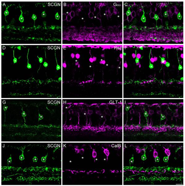
Figure 3.
SCGN is localized to DB1 bipolar cells. Confocal projections of vertical sections labeled for SCGN (left panels) and various OFF bipolar cell markers (middle panels). Merged images are shown in the right-hand panels. A–C: Intensely labeled SCGN-positive bipolar cells (asterisks) lack Gαo staining, indicating that they are likely OFF cone bipolar cells (projection of 7 × 0.4 μm). D–F: SCGN-positive bipolar cells (asterisks) do not colocalize with the flat midget bipolar cell marker, recoverin (projection of 7 × 0.9 μm). G–I: Staining for SCGN and GLT-1, a marker of DB2 and FMB bipolar cells. SCGN-positive bipolar cells (asterisks) show weak GLT-1 immunoreactivity; however, the more intensely stained DB2 axon terminals are in a more proximal position in the IPL (projection of 3 × 1.0 μm). J–L: Staining for SCGN and the DB3 marker, calbindin. SCGN-positive bipolar cells (asterisks) lack calbindin immunoreactivity (6 ×0.9 μm). All images are from peripheral retina except D–F, which is taken from mid-peripheral retina. Scale bar = 10 μm. [Color figure can ≈be viewed in the online issue, which is available at wileyonlinelibrary.com.]

