Figure 5.
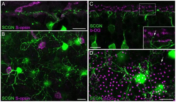
DB1 bipolar cells frequently contact S-cones but rarely contact rods. A: Confocal projection (3 × 1.0 μm) of a peripheral retinal section stained for SCGN and S-opsin showing that SCGN-labeled bipolar cell dendrites contact two S-cone pedicles. B: A single confocal section showing double labeling of SCGN and S-opsin in whole-mounted mid-peripheral retina. SCGN-labeled DB1 bipolar cells nonselectively contact M/L cones and S-cone pedicles within their dendritic arbors. C,D: Double labeling of SCGN and β-dystroglycan (b-DG, as a marker for rod terminals and cone pedicles) on a vertical section (C, single confocal image) and whole-mount (projection of 2 × 1.0 μm) from mid-peripheral retina. Rare examples of SCGN-positive dendrites that appeared to contact rod terminals are indicated (arrows). Scale bars = 10 μm. [Color figure can be viewed in the online issue, which is available at wileyonlinelibrary.com.]

