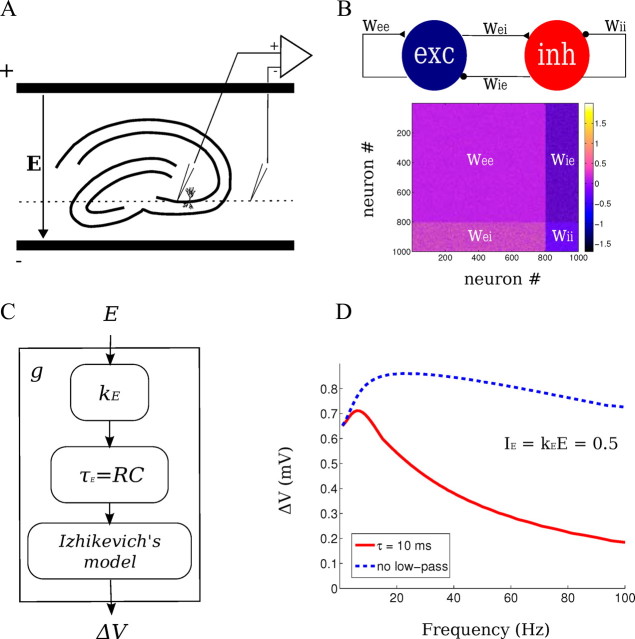
Figure 1.
Experimental and computational network methods. A, Experimental setup: spatially uniform electric fields were applied across hippocampal slices in an interface chamber. Recording of LFPs and intracellular potentials were performed in the CA3 region of hippocampus relative to an isopotential electrode in the bath (dotted line). The orientation of the pyramidal layer of the CA3c region was perpendicular to the applied field. B, Computational network model: 800 excitatory and 200 inhibitory neurons are synaptically connected with various synaptic strengths w. C, The electrical stimulation in the model was implemented as low-pass-filtered current (τE = 10 ms) that is proportional to the applied field E. D, The polarization of the single-neuron model of Izhikevich to a current injection (dashed blue line) exhibits low-pass characteristics. A better match to experimental data is observed with the additional low-pass filter. The combined model (solid red line) has a peak gain at ∼7 Hz.

