Fig. 4.
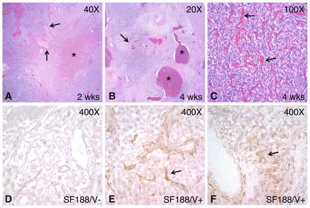
Microscopic and immunohistochemical xenograft analysis. a A small but cellular SF188/V+ tumor in the left side of the panel contains dilated vessels (arrows) extending into host brain with few or no neoplastic cells on the right. A focus of hemorrhage is also present (asterisk). b A larger xenograft removed after 4 weeks of growth contains a number of massively dilated vessels (asterisks) with focal thrombosis. Vessels with lesser degrees of dilatation are also present (arrow). c In addition to vascular dilatation, proliferation of smaller vessels resulted in a dense network throughout most of the tumor (arrows). d–f Only weak VEGF immunoreactivity was present in SF188/V− cells, while SF188/V+ cells gave rise to xenografts with moderate to strong VEGF expression in and around vessels (arrows)

