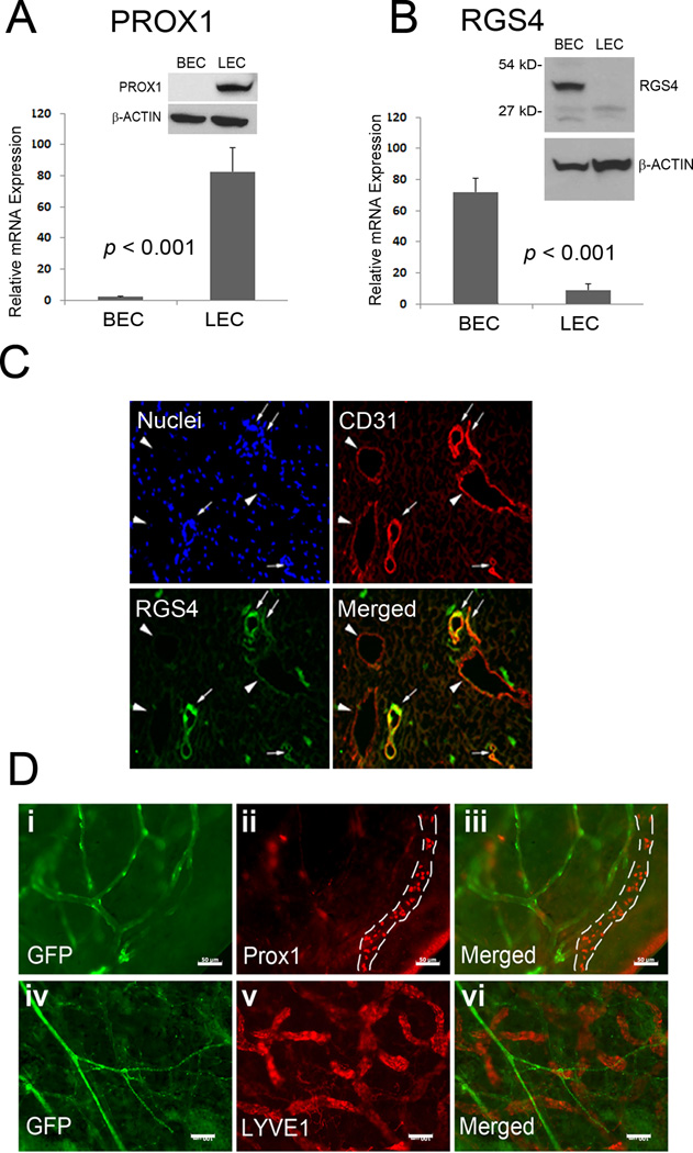
Figure 2. RGS4 is predominantly expressed in BECs, compared to LECs.
Expression of PROX1 (A) and RGS4 (B) in human primary dermal BECs and LECs was determined by qRT-PCR and western blot analyses. Data are shown as average relative expression ± standard deviation. qRT-PCR and western blot analyses were repeated at least two times. (C) Immunofluorescent analyses with DAPI (Nuclei), anti-CD31, and anti-RGS4 antibodies on neonatal human foreskin section show that RGS4 is mainly expressed in CD31-high blood vessels (arrows), but not in CD31-low lymphatic vessels (arrowheads). Scale bar, 20 µm. (D) Whole mount staining of the ears of RSS4-GFP adult mice for two lymphatic markers Prox1 (i–iii) and LYVE-1 (iv–vi). (i, iv) GFP expression from transgenic RGS4-GFP mice; (ii, v) antibody staining for Prox1 and LYVE1, respectively; (iii, vi) merged images. A Prox1-positive lymphatic vessel was marked with dotted lines in panel (ii, iii). Scale bars: 50 µm (i–iii) & 100 µm (iv–vi). Immunofluorescent analyses were performed more than twice with consistent results.

