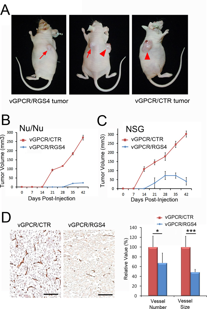
Figure 6. RGS4 inhibits vGPCR-induced tumor formation.
(A) Tumor formation by SVECs expressing vGPCR alone (vGPCR/CTR) or vGPCR and RGS4 (vGPCR/RGS4) was evaluated in athymic nude mice. Arrow indicates vGPCR/RGS4 tumor and arrowhead indicates vGPCR/CTR tumor in the same mouse at 6 weeks post subcutaneous inoculation. (B,C) Growth curves of vGPCR/CTR and vGPCR/RGS4 tumors in athymic nude (Nu/Nu) (B) or NOD-SCID IL2Rγnullmice (NSG) (C). Equal numbers of vGPCR/CTR and vGPCR/RGS4 cells were subcutaneously injected at the right and left flank area, respectively, five female mice per group (n=5). Experiments were performed twice with similar results and tumor volume is shown as average tumor volume ± standard deviation of one representative experiment. (D) Reduced tumor-associated angiogenesis by RGS4-expressing tumors. Sections prepared from paraffin-embedded tumors harvested from athymic nude mice at Day 42 were subjected to immunohistochemistry analysis for CD31. Number and size of CD31-positive vessels were analyzed using the NIH ImageJ program and are displayed as relative value ± standard deviation. Scale bars, 100 µm; *, p <0. 05; ***, p <0. 001. Animal studies were performed three times with consistent results.

