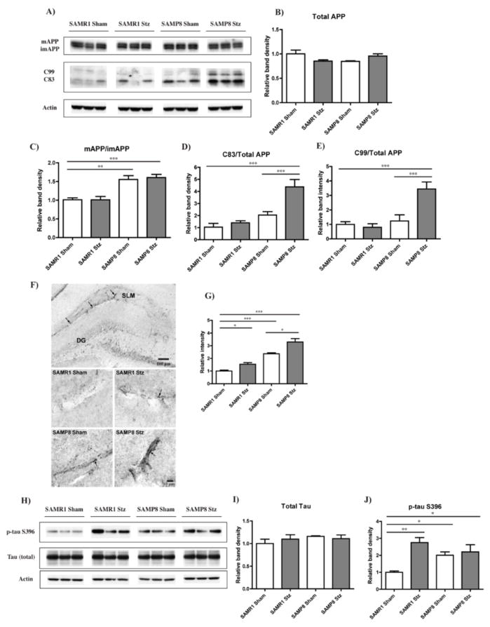
Figure 3. AD-related pathology in the hippocampus of T1D mice.
T1D SAMR1 and SAMP8 mice were sacrificed four months after administration of Stz, and their brains were removed and processed for both Western blot (WB) and immunohistochemistry (IHC). (A) WB analysis of APP processing in RIPA-soluble fraction of hippocampal tissue, using an antibody against the C-teminus of APP. Full length APP, both its mature (mAPP) and immature (imAPP) forms, and the APP cleavage products C99 and C83 were detected. Actin was used as a loading control. (B) Quantification of the levels of total APP (mAPP+imAPP) in relation to actin. (C) A significant increase in mAPP relative to imAPP in both the SAMP8 Sham and T1D mice was identified, along with elevated levels of C99 (D) and C83 (E) only in the T1D SAMP8 mice. **p<0.01, ***p<0.001, One-way ANOVA followed by Newman-Keuls post-hoc test (n = 5–6/group). (F) IHC analysis of Aβ in the hippocampus identified blood vessel shaped-like reactivity, particularly noticeable in the stratum lacusonum moleculare (SLM). Original magnifications: x100 (first panel, SAMP8 T1D), x630 (rest of the panels). (G) Quantification of Aβ revealed increased reactivity (arrows) in SAMP8 mice. T1D increased Aβ aggregation in both the SAMR1 and the SAMP8 mice. *p<0.05, ***p<0.001, One-way ANOVA followed by Newman-Keuls post-hoc test (n = 3/group). (H) WB analysis of total tau and tau phosphorylated at Ser396 (p-tau S396) in RIPA-soluble fractions of hippocampal tissue. No changes in total tau levels, relative to actin, were identified between the groups (I); however, T1D SAMP8 and Sham mice and T1D SAMR1 mice showed significantly higher levels of p-tau S396 in comparison with the SAMR1 Sham mice (J). *p<0.05, **p<0.01, One-way ANOVA followed by Newman-Keuls post-hoc test (n = 4–5/group). All data are mean ± SEM.

