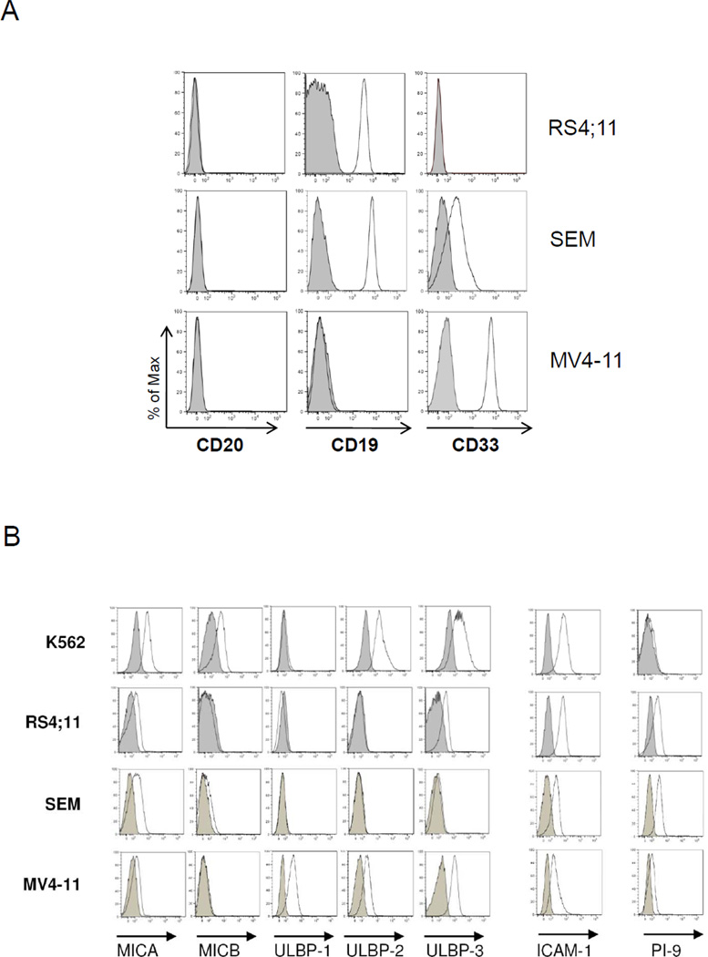
Figure 1.
Characterization of MLL-rearranged cell lines by flow cytometric analyses. Expression of (A) CD20, CD19, and CD33; (B) NKG2D ligands (including MICA, MICB, ULBP-1, ULBP-2, and ULBP-3), adhesion molecule ICAM-1, and intracellular PI-9. Standard NK cell target K562 was used as a positive control for comparison. Data are representative of three independent experiments. Shaded histograms represent the isotype control.

