
Figure 1.
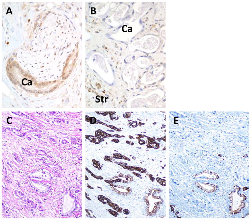
Deviant SMAD4 and CDKN2A immunohistochemical labeling patterns in pancreatic cancer tissues. A. Intact Smad4 immunolabeling in a primary carcinoma. Both nuclear and cytoplasmic labeling for SMAD4 is present within the neoplastic glands (Ca) in an area of perineural invasion. B. Loss of SMAD4 immunolabeling in a liver metastasis derived from the carcinoma shown in A. In this example, no labeling of SMAD4 is seen within the neoplastic glands (Ca). By contrast, positive labeling of surrounding stromal cells (Str) is present. C. Hematoxylin and eosin stained section of infiltrating pancreatic carcinoma. D. CK19 labeling of the carcinoma shown in C indicating strong positive labeling throughout the neoplastic epithelium. E. Example of focal loss of CDKN2A immunolabeling in the carcinoma shown in C. No labeling of CDKN2A is seen in the neoplastic epithelium within the upper half of the shown section, whereas strong positive labeling is seen within scattered neoplastic glands in the lower half.