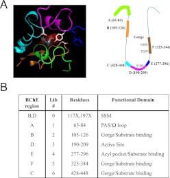
Fig. 1.
A depiction of mutation libraries of hBChE. A, depiction of each designed mutation library around the active site of hBChE. Left, three-dimensional view of the active site of hBChE. Right, a cartoon model showing the active site of hBChE. Each color represents a corresponding peptide sequence that was covered by each individual library: green, library 1; orange, library 2; pink, library 3; blue, library 4; brown, library 5; aqua, library 6. B, list of libraries and their corresponding function. Library 0 is a site-saturated mutation library. Libraries 1 to 6 are randomized mutation libraries designed to cover six structurally relevant regions around the active pocket of hBChE.

