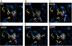
Fig. 7.
Structural models of hBChE variants. Three-dimensional models of six hBChE variants identified in the molecular evolution screen and characterized by kinetics. A, wild-type hBChE. B, G117H. C, G117R. D, G117N. E, L125V. F, E197C. The catalytic triad of the hBChE substrate binding domain is shown as a yellow backbone on the left in every image. Substrate is shown as gray in the center. The amino acid that underwent mutation is shown with a white backbone near the bottom right corner (B–D); right middle (E), and bottom left (F). Hydrogen bonds are shown as dashed lines.

