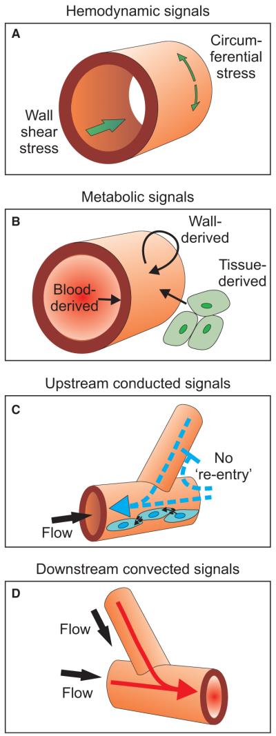
Fig. 1.
Schematic diagram showing haemodynamic forces and other factors acting on vessel walls. (A) Haemodynamic signals include wall shear stress acting on endothelial cells because of blood flow and circumferential stress generated in the wall as a result of intravascular pressure. (B) Metabolic signals may be generated by the blood, the wall and the surrounding parenchymal cells, and may act on all components of the wall. (C) Upstream propagation of signals occurs via conducted responses propagated along the vascular walls. Cell-to-cell coupling by gap junctions mediates this process. The blunt-headed arrow represents the no-re-entry phenomenon (see text for explanation.) (D) Convective transport of vasoactive metabolites provides a mechanism for downstream propagation of signals.

