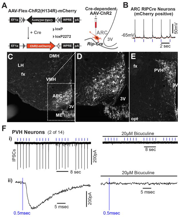
Figure 6. Projection of ARC RIP-Cre Neurons.
(A) Diagram of AAV-Flex-ChR2(H134R)-mCherry (left) and schematic indication of the stereotaxic injection into the ARC of Rip-Cre transgenic mice (right).
(B) Representative voltage tracing showing light-driven spikes in a current-clamped arcuate neuron marked by mCherry fluorescence. Blue tickmarks represent 0.5msec light flashes at 0.5Hz.
(C–E) Immunohistochemistry for mCherry in the hypothalamus of virus-injected Rip-Cre transgenic mice. mCherry-expressing RIP-Cre neurons in the ARC are shown in (C) and in a zoomed view in (D). (E) mCherry-expressing RIP-Cre neuron fibers in the PVH. DMH: dorsomedial hypothalamus; VMH: ventromedial hypothalamus; ME: median eminence; ARC: arcuate nucleus; SCN: suprachiasmatic nucleus; PVH: paraventricular hypothalamus; 3V: third ventricle; fx: fornix; opt: optic tract.
(F) - i) Light-evoked IPSCs in a PVH neuron before (left) and after (right) the addition of 20μM bicuculine in response to clusters of light pulses. Blue tickmarks represent 0.5msec light flash at 0.5Hz. ii) Zoomed in view of response to a single pulse of light.
See also Figure S6.

