


Figure 2.
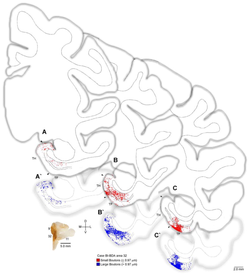
Distribution of terminations from ACC to the parahippocampal cortices. Rostral (A) through caudal (C) tracings of coronal sections through parahippocampal cortices (areas TH, TF) of the rhesus monkey brain (Case BI) show the areal and laminar distribution of small (red) and large (blue) labeled boutons following injection of the anterograde tracer BDA in ACC area 32 (coronal section lower left). Double lines in the middle of the cortex denote layer IV. Abbreviations: ACC: anterior cingulate cortex; BDA: biotinylated dextran amine.