


Figure 3.
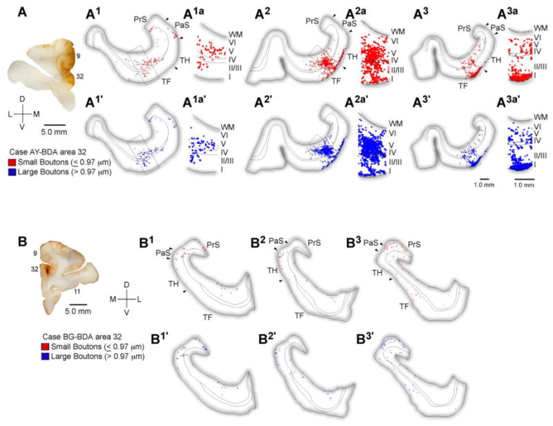
Distribution of terminations from ACC to the parahippocampal cortices. A, Injection site in ACC area 32. A1-3a, Rostral (A1) through caudal (A3) tracings of coronal sections through the parahippocampal areas TH and TF show the areal and laminar distribution of small (red) and large (blue) labeled boutons following injection of the anterograde tracer BDA in ACC area 32 (A, case AY) and the adjacent part of medial area 9. Higher magnification insets show the nearly even laminar distribution in the superficial and deep layers. B, Rostral through caudal tracings of coronal sections through the rhinal and parahippocampal cortices following a focal injection in area 32 (B, case BG). Abbreviations: ACC: anterior cingulate cortex; BDA: biotinylated dextran amine; PaS: parasubiculum; PrS: presubiculum; WM: white matter.