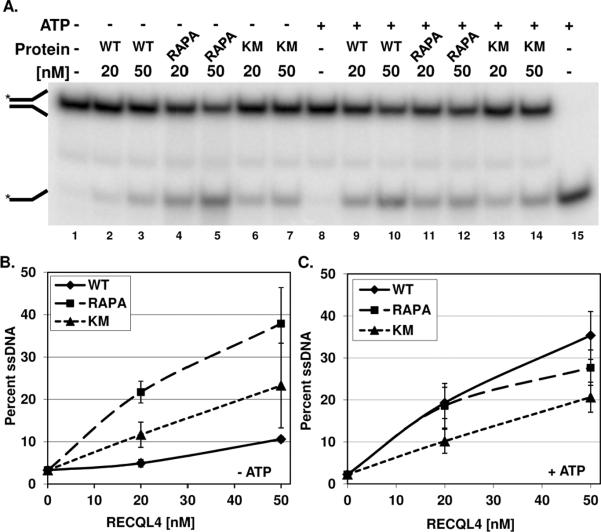
Figure 4. Strand separating activity in the presence of excess single stranded DNA.
The strand separating activity of WT and RAPA RECQL4 was determined using the labeled forked duplex B1/T1 (0.5 nM) in the presence of 12.5 nM cold B1 as described in the Materials and Methods. A, a representative phosphorimage of a gel displaying the strand separating activity of WT (lanes 2–3 and 9–10), RAPA RECQL4 (lanes 4–5 and 11–12), and K508M RECQL4 (lanes 6–7 and 13–14) in the absence and presence of ATP is shown. The Δ symbol denotes the heat denatured DNA substrate. The position of the single stranded and forked dsDNAs are denoted by the images on the left side of the gel. B and C, graph of the quantification of three independent gels each in the absence and presence of ATP, respectively. The mean and standard deviation are reported.

