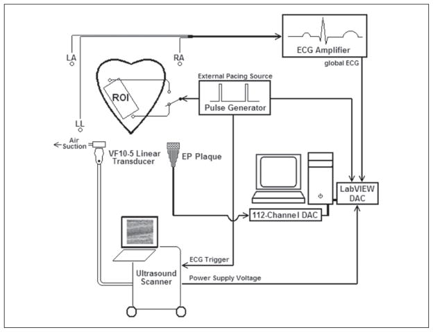
Figure 1.
Graphical representation of the experimental setup. An exposed and externally paced canine heart is imaged with a transducer/vacuum apparatus placed over a region of interest (ROI) directly on the left ventricular free wall. Electrocardiogram (ECG)–gated B-mode and acoustic radiation force impulse images, the voltage from the power supply of the ultrasound scanner, global ECG, and stimulating pulse waveforms were simultaneously recorded via data acquisition cards (DACs) and temporally registered. The procedure was performed while pacing on either side of the transducer. The transducer/vacuum apparatus was then removed and replaced with a 112-electrode electrophysiology (EP) plaque. The procedure was then repeated while recording local epicardial potentials over the same ROI.

