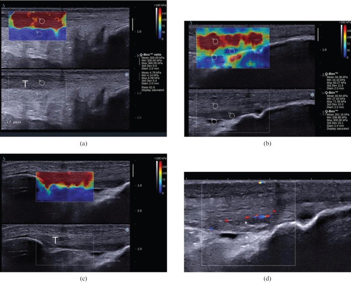
Figure 3.
Longitudinal shear wave elastograms of a normal (a) Achilles and (c) patella tendon, as well as (b, d) a case of distal patella tendinopathy in a 23-year-old football player. The elasticity qualitative and quantitative scale is presented at the upper right corner of the images. Measurements (mean, minimum, maximum and standard deviation) within the circular region of interest (ROI) are presented in kilopascals ranging from 0 (dark blue) to 300 (dark red). (a, c) The normal Achilles and patella tendons (T) appear as homogeneous stiff (red) structures, as opposed to fat, which is homogeneously soft (blue). (a) The mean stiffness of a representative area at the mid-portion of the Achilles free tendon is 300 kPa. (d) In the case of distal patella tendinopathy, the tendinopathic area appears hypoechoic with neovascularity (asterisk). (b) In the corresponding elastogram, the abnormal area appears softer (blue; mean elasticity 40.94 kPa) compared with the stiffer normal tendon (red; mean elasticity 261.16 kPa). The small amount of fluid in the deep infrapatella bursa appears softer than the tendinopathic area (blue, mean elasticity 34.38 kPa).

