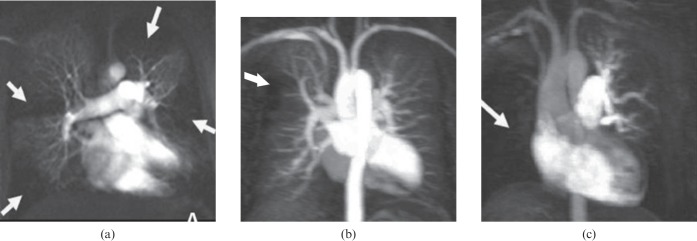
Figure 9.
Perfusional aspect in chronic thromboembolic pulmonary hypertension. Maximum intensity projection images of a single perfusion phase obtained with contrast-enhanced MR-derived pulmonary perfusion in three patients: (a) multiple perfusion defects (white arrows), (b) focal perfusion defect in the anterior segment of the right lung upper lobe (white arrow) and (c) complete perfusion defect in the right lung (white arrow).

