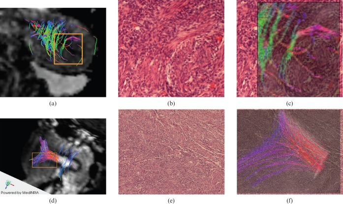
Figure 4.
3 T MR-diffusion tensor imaging (DTI) reconstruction was compared with the corresponding histological specimens of the ex vivo uteri that accurately show similar fibre architecture for both circular (first row) and longitudinal (second row) fibre bundle. (a) Circular fibre bundle superimposed to DTI post-processed image. (b,c) Histological specimen (haematoxylin and eosin) without and with circular fibre superimposition. (d) Longitudinal fibre bundle superimposed to DTI post-processed image. (e,f) Histological specimen (haematoxylin and eosin) without and with circular fibre superimposition.

