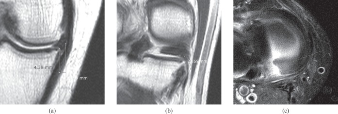
Figure 4.
A 59-year-old male with knee joint swelling. (a) Proton density coronal image (repetition time, 3000 ms; echo time, 35 ms) of the knee at the mid-point shows extrusion of the medial meniscus with a distance of 4.39 mm. (b) A proton density coronal image (repetition time, 3000 ms; echo time, 35 ms) 15 mm behind (a) shows a 27.7 mm transverse meniscus length. The meniscal extrusion transverse ratio was 0.16. Complete disruption of the medial meniscal root is seen. (c) Fat-saturated proton density imaging (repetition time, 3000 ms; echo time, 36 ms) reveals a definite tear of the posterior root of the medial meniscus. The posterior root tear was confirmed arthroscopically.

