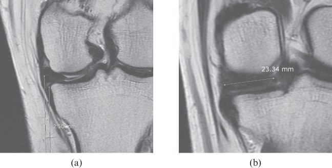
Figure 7.
A 42-year-old female with knee joint pain lasting for 3 months. (a) A proton density coronal image (repetition time, 3000 ms; echo time, 35 ms) of the knee at the mid-point shows extrusion of the medial meniscus with a distance of 2.50 mm. (b) A proton density coronal image (repetition time, 3000 ms; echo time, 35 ms) 15 mm behind (a) shows a 23.3 mm transverse meniscus length. The meniscal extrusion transverse ratio was 0.11. A posterior root tear of the medial meniscus was diagnosed on arthroscopy.

