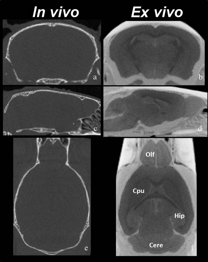
Figure 5.

In vivo and ex vivo Micro-CT images. Three-plane images of (a, c, e) in vivo and (b, d, f) ex vivo micro-CT studies. The ex vivo images show clearer signal differences between the grey matter, the white matter and the ventricular spaces than the in vivo images. (a, b) axial, (c, d) horizontal and (e, f) sagittal sections. Brain structures including olfactory bulb (Olf), hippocampus (Hip), caudate putamen (Cpu) and cerebellum (Cere) are readily distinguishable.
