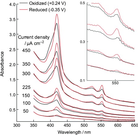
Figure 1.

Absorbance spectra of G. sulfurreducens biofilms while growing at oxidizing (+0.24 V) potentials (black traces). After collecting each scan, electrodes were poised at −0.35 V for 10 min to obtain data from the fully reduced state (red trace). Application of potentials as high as +0.5 V did not further oxidize cytochromes during growth. Inset expands data from 50–225 μA cm−2. All potentials are versus standard hydrogen electrode (SHE).
