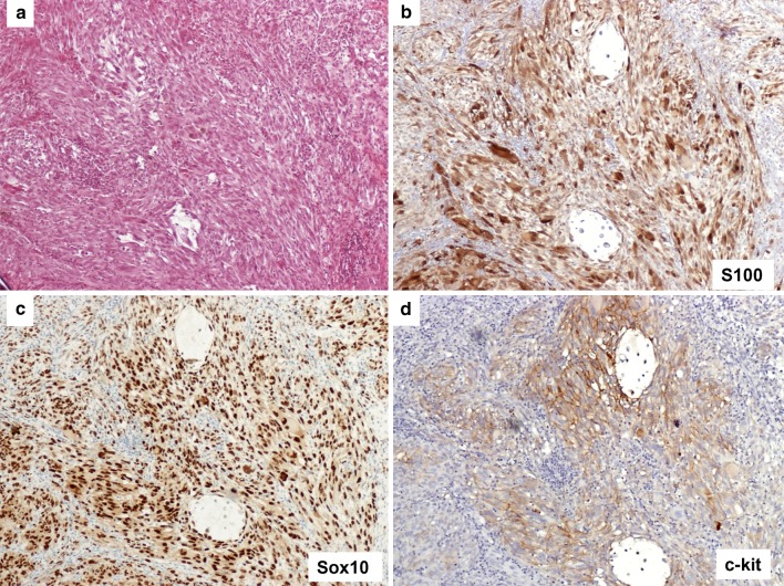
Fig. 3.
Representative case of mucosal melanoma, spindle cell morphology. a Section of melanoma composed of spindle shaped tumor cells (H&E). b Immunostain for S100 shows nuclear and cytoplasmic staining. c Sox10 highlights all nuclei of the melanoma cells. d Immunostain for c-kit shows membrane staining (3+). Original magnification ×100 for a–d

