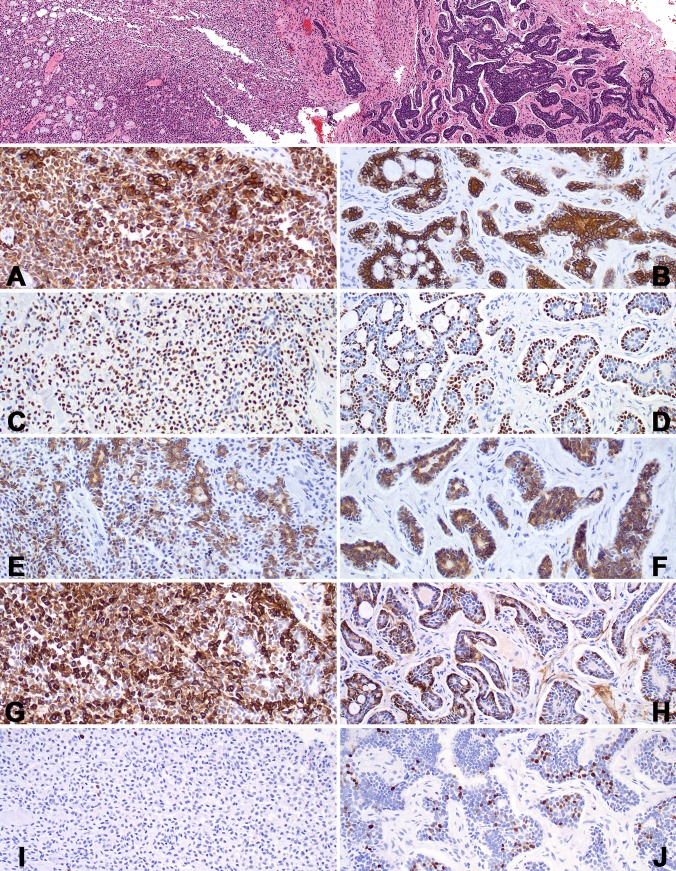
Fig. 7.
A lower power field shows a pleomorphic adenoma (left) transitioning into adenoid cystic carcinoma (right). The immunohistochemistry reactions are shown for the pleomorphic adenoma (left) and the adenoid cystic carcinoma (right) in each row. A and B: CK7: Note the luminal cell accentuation in the ACC. C and D: p63: There is a basal accentuation in the ACC. E and F: CD117. G and H: SMMHC: There is variable accentuation in the ACC. I and J: Ki67. Note the number of positive nuclei in the ACC in comparison to the PA

